Category: E-Report
To Our Customers and Partners, Thank You.
It’s the time of year to reflect, celebrate and give thanks for so many things. This COVID-19 dominated year has certainly impacted all of us. At Epica Animal Health, we would like to extend to our customers and partners our deepest gratitude. We are so grateful to our community of existing and prospective HDVI CT platform users for your sincere interest, ongoing support and daily practice usage of our ultra-high resolution imaging platform. We are grateful to have the opportunity to empower veterinarians across the nation to see and discover more and deliver faster and more accurate diagnosis to the #furryfriends that we call #family.
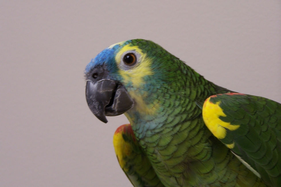
HDVI - Avian and Exotics
During this incredibly challenging period, we are awed by your continuous commitment to the treatment and care of our #pets, #emotionalsupportanimals, #wildlife and #exoticanimals everywhere. This is why Epica™ remains committed to enabling your #continued education! As a token of our appreciation, the #HDVI team would like to present you with this collection of 9 HDVI Avian and Exotic case studies from Mobile Pet Imaging, Scarlet Imaging and Parrish Creek Veterinary Hospital. Start your commitment to #continuededucation today by clicking the link below and accessing this complimentary interactive E-Report. #thanksgiving2020
This multimedia collection of Avian and Exotics HDVI Case Studies that were submitted by Vimago™ users M. Scott Echols, DVM, Diplomate ABVP - Avian Practice from Parrish Creek Veterinary Clinic and Xavier Meaux, DVM from Mobile Pet Imaging. This book includes CT reports and ultra-high resolution images from an array of avian and exotic animals: African Grey, Andean Condor, Pelican, Rat, Bamboo Shark, Blue and Gold Macaw, Blue Fronted Amazon Parrot, Bufflehead Sea Duck, and Cockatiel.
Avian & Exotics HDVI Applications:
Hard Tissue Applications:
Skulls — sinus, nasal, bulla, ears, brain, oncology, trauma
Orthopedics — stifles, elbows, hips, carpus/tarsus, angular limb deformities, OCD, cartilage lesions, fractures
Dentistry
Spine — cervical, thoracic, lumbar
Soft Tissue Applications:
Tumors, foreign bodies
Thorax — lungs, heart
Blood vessels, met checks
Abdomen — liver, pancreas, kidneys, adrenals, GI, shunts, oncology
Angiography
Diagnostic Fluoroscopy — coughing, swallow studies, UG studies, shunts, collapsing tracheas
Interventional Fluoroscopy – biopsies, endoscopy guidance, stents, SUB.
Surgical planning, guided biopsies, and minimally invasive surgeries

Radiographs and ultrasound can sometimes indicate that anomalies or irregular pathology may exist within a patient but, too often they go unnoticed and untreated. However, Vimago™ HDVI allows veterinarians to see the exact size, scope and location of the masses, fractures or lesions, all in one scan. The results can often be viewed in 3D to provide a more accurate understanding of the patient’s prognosis, but can also be viewed as a 2D Radiograph or Live Fluoroscopic video for swallow studies and for the identification of gastrointestinal (GI) and respiratory issues.

Vimago™ Customer Spotlight:
All radiology report findings and treatment recommendations in this collection were provided by Dr. Scott Echols.
About Dr. Echols

Dr. Echols graduated from the Texas A&M College of Veterinary Medicine in 1995 and completed his residency in avian medicine and surgery at the Medical Center for Birds in Oakley, California in 1999. In addition to being a frequent internationally recognized author and lecturer, Dr. Echols has been awarded the TJ Lafeber International Avian Practitioner of the Year and Texas Non-traditional Species Practitioner of the Year Awards. Dr. Echols serves as an Adjunct Professor for Texas A&M College of Veterinary Medicine as well as a Visiting Professor for many universities in the US and abroad. Dr. Echols is a past president of the Association of Avian Veterinarians, a reviewer of scientific papers and has worked with numerous veterinary organizations in different capacities.
While Dr. Echols works internationally, he and his family live in Utah and he most frequently works at Parrish Creek Veterinary Clinic.
About Parrish Creek Veterinary Clinic

Parrish Creek Veterinary Clinic is one of the few veterinary hospitals that specializes in the care of birds. There are only three Avian Medicine and Surgery doctors in Utah recognized as specialists, and we have them all here! Dr, Antinoff, Dr. Folland and Dr. Echols are diplomates of the American Board of Veterinary Practitioners. They have considerable experience in the care of these unique pets and understand their special needs. You can have confidence we will provide the expertise and knowledge necessary to keep your bird or other exotic pets healthy.
We are members of the following associations which gives us access to the most current information on the care of exotic species.
In addition, we spend several hours each year in continuing education to stay on the cutting edge of care for birds and exotic pets.
Please visit our pet care information page for care sheets for many different species.
Please visit our website at: parrishcreekvet.com
The African Grey Case was prepared for Mobile Pet Imaging by Dr. Scott Echols
About Mobile Pet Imaging

What is Mobile Pet Imaging?
Mobile Pet Imaging is a veterinary service that offers the latest in high-definition CT Scans and fluoroscopy—in a mobile unit that comes to vets’ offices.
What is a CT Scan?
CT stands for Computed Tomography. It is basically a series of very fine X‑rays that the computer then assembles into a 3D image. CT scans are especially helpful in evaluating the skull, brain, sinuses, inner ear, eye sockets, spine and discs, bones, joints, and soft tissues.

How are we different?
Mobile Pet Imaging HD CT Scans offer more resolution than traditional diagnostic imaging tools.
In most cases, Mobile Pet Imaging’s CT scans are more affordable than traditional CT Scans.
Mobile Pet Imaging comes to veterinarians’ offices, so there is no need for referral to a different hospital or office. Your pet will be scanned in the mobile unit at your veterinarian’s office and returned to them for aftercare. A licensed veterinarian is with your pet at all times during our service.
A CT Report by a board-certified radiologist is emailed to the primary veterinarian within 24 hours (can be ordered “rush” if needed).

Where are we located?
Mobile Pet Imaging provides services from a custom-built, state-of-the art mobile unit specifically designed to safely transport high-tech equipment and provide a safe, clean and tranquil place to perform the procedures.
The Mobile Pet Imaging mobile unit services the Miami-Dade, Broward, Palm Beach and Monroe counties, but can travel outside our standard service area upon request.
Some Interesting Cases.
Mobile Pet Imaging provides services to more than just dogs and cats. In the first year of business, Mobile Pet Imaging has had the honor of helping diagnose all sorts of animals: from a pet squirrel, a cockatoo and a Komodo dragon, to rabbits and a marmoset.
Mobile Pet Imaging’s equipment accommodates animals up to 200 lbs., including the chimpanzee scanned for the Lion Country Safari in Palm Beach County.
A case done at the Palm Beach Zoo caught the eye of local news; two recent cases at a private practice will be featured on a national TV show.
Mobile Pet Imaging is also proud to have done many discounted scans for rescue groups, veterinarians and their staff, zoos and wildlife facilities throughout South Florida.
BACKGROUND INFO
Mobile Pet Imaging was conceived by Dr. Pedro Armstrong, a veterinary internist in South Florida, who wanted to bring the latest in CT technology to ALL patients, in a more convenient and affordable manner than was available. In 2014, Dr. Armstrong partnered with Dr Meaux to create Mobile Pet Imaging.
Dr. Pedro Armstrong DVM, DACVIM- Dr. Pedro Armstrong DVM, DACVIM- Managing partner and internal medicine specialist, has been practicing in the Miami area since 1996. In 2005, Dr. Armstrong is also one of the founders and a previous owner and internist at Southeast Veterinary Referral Center and the Pet Emergency Room in Miami.
Dr. Xavier Meaux- Partner and Lead Mobile Unit Veterinarian.
Dr. Ana V. Caceres, DVM, DACVR- Head of Diagnostic Imaging at University of Pennsylvania School of Veterinary Medicine and consulting radiologist for Mobile Pet Imaging.
Please visit our website at: itstimetoseemore.com
Comments